ImmYOUnology

More than just vaccines


1 Comment

Allergies are no fun…but the biology behind them is!

Spring is nearly upon us and along with trees and flowers, seasonal allergies will bloom once again.  Even though allergies can be annoying, debilitating and even life-threatening, the science behind them is fascinating.  Science published a timely paper at the end of February describing some of the ways different kinds of allergens work.  Allergens are small parts—individual proteins or molecules—of things that cause allergic responses.

The group who published the study worked with cells called mast cells, one of the common types of the immune cells that respond to allergens and make you itchy, sneezy and swollen.  Before they can activate mast cells, allergens have to be recognized by a particular type of antibody, or immunoglobulin, called immunoglobulin E, or IgE.  On one end, IgE binds an allergen, and on the other it interacts with a protein receptor on mast cell surface.

By connecting the mast cell to the allergen, IgE gives the mast cell permission to do its thing, and its thing is called degranulation.  Mast cells are brimming with packets, or granules, of histamine and heparin and other proteins that damage microbes as well as tissue.  When the cells degranulate, they open up and release their contents into whatever tissue they happen to be in—the skin, the lungs or the gut for example.  Many of the contents released make blood vessels leaky and attract lots of immune cells, causing inflammation.  Antihistamines prevent the released histamine from binding its receptors on blood vessel cells.  Another treatment option currently under investigation is a drug that blocks the interaction between IgE and the receptor on mast cells to prevent this process from even getting started.  

The recent Science paper took a close look at the mast cell response to IgE-bound allergen and showed just how fine-tuned it can be. The researchers activated mast cells with allergens that bound tightly or weakly to IgE and found that the strength of the interaction, also called affinity, changed the way that mast cells responded.

http://commons.wikimedia.org/wiki/File%3ASMCpolyhydroxysmall.jpg

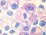
Skin mast cells stained with Toluidine blue

The researchers could study mouse mast cells in culture dishes, because mast cells grow up from stem cells inside bone marrow.  So they grew up mast cells from mouse bone marrow and then gave them the strongly binding allergen (high affinity) or the weakly binding one (low affinity). They could get the mast cells to respond and degranulate with both, but it took 100 times more of the weak binding allergen to get the same response caused by the strong one.

To understand how allergic reactions work in living creatures, researchers often sensitize mouse ears by exposing them to an allergen and later re-introduce the allergen through the bloodstream. Then they can measure how inflamed the ears get and how many and what kinds of immune cells travel to the ear after injecting the allergen.  In this study, the strong binding allergen caused more intense and more sudden ear inflammation and immune cell infiltration than the weaker binding allergen.

So how does this fascinating mechanism actually relate to human allergies, which for some people is a life-threatening condition.  Although some allergies go away with age, there is currently no permanent cure for those that don’t.  Treatment of serious allergies is centered around desensitization immunotherapy, which is just repeated exposure to small doses of allergen over time.  The treatment may last anywhere from months to a lifetime and there are no biomarkers, or biological tests, that tell doctors when the treatment is working.  Instead, they simply test allergens on patients, which could mean pricking the skin or making them eat peanuts one at a time until they do or don’t get sick.  

A clinical study that came out in January helped me understand how knowledge of allergen binding strength could be helpful in treatment.  In this study, children with milk allergies were undergoing oral immunotherapy, which in this case simply meant they had to drink small amounts of milk that were increased over time.  The researchers collected serum samples from the kids in the study and measured levels of IgE as well as the affinity of IgE for proteins found in cow’s milk to see if either would change as kids became more tolerant to milk.

In some cases, the immunotherapy had to be discontinued because the reactions to milk were too severe.  The researchers found that the IgE from the kids whose treatment was discontinued bound more tightly to milk proteins compared to kids who responded well to the treatment.  So the strength of the interaction between IgE and allergens does matter, at least in the case of cow’s milk allergies. This study didn’t look at mast cells, but it does indicate that the molecular details of how IgE connects allergens to mast cells are worth studying.  Those details can provide clues about what is going on inside a person with allergies and how well they may respond to immunotherapy.

Sources:

Mastcellaware.com (A whole website about Mast Cells)

Suzuki R., Leach S., Liu W., Ralston E., Scheffel J., Zhang W., Lowell C.A. & Rivera J. (2014). Molecular Editing of Cellular Responses by the High-Affinity Receptor for IgE, Science, 343 (6174) 1021-1025. DOI:

Savilahti E.M., Kuitunen M., Valori M., Rantanen V., Bardina L., Gimenez G., Mäkelä M.J., Hautaniemi S., Savilahti E. & Sampson H.A. & (2014). Changes in IgE and IgG4 epitope binding profiles associated with the outcome of oral immunotherapy in cow’s milk allergy, Pediatric Allergy and Immunology, n/a-n/a. DOI:

Moran T.P., Vickery B.P. & Burks A.W. (2013). Oral and sublingual immunotherapy for food allergy: current progress and future directions, Current Opinion in Immunology, 25 (6) 781-787. DOI:


1 Comment

Tuning down the immune system could improve the flu vaccine

This post is based on an article I recently wrote for an internship application, so it’s more formal than a typical post, but I think it’s a cool story that helps explain how the flu vaccine works. Enjoy! And stay healthy!

After more than 2,000 confirmed cases and over twenty deaths, the 2013-14 flu season is still approaching its peak.  Vaccination remains the best prevention despite the flu vaccine’s hit-or-miss reputation.   Each year the U.S. Food and Drug Administration recommends three strains of influenza that the World Health Organization believes are worth targeting, and six months later the season’s new vaccine is distributed.

influenza-virus-labels

This nasty viral particle is trying to get inside a cell. It’s covered in NA (red) and HA (blue) proteins.

One of the flu vaccine’s biggest problems is its inability to induce immunity against multiple viral subtypes.  Subtypes of the influenza A virus, like H1N1 or H5N1 are distinguished by the surface proteins hemagglutinin (HA) and neuraminidase (NA).  The vaccine can protect against a few subtypes at a time, but if a subtype not included in the shot makes a strong appearance one season, not much can be done to prevent it from spreading.  This year, the vaccine is pretty spot on. It includes H1N1 which has been making a comeback this year.

This problem has driven researchers to pursue a universal vaccine that could protect against multiple subtypes.  This type of protection is called heterotypic immunity.  One group of scientists from St. Jude Children’s Research Hospital hit on an unexpected way to expand the reach of one flu vaccine to multiple subtypes.  Dr. Maureen McGargill and her group published their study in Nature Immunology in December.  They studied how a common immunosuppressive drug called rapamycin influenced the ability of vaccinated mice to generate heterotypic immunity.  They vaccinated mice with one viral subtype and infected them with three other lethal subtypes.  Surprisingly, the mice who got rapamycin were better able to resist infection by all the subtypes, including an altered H5N1 strain, commonly known as the avian flu.

Rapamycin is commonly used to dampen the immune system to prevent organ transplant rejection.  It blocks an immune system regulating protein called mTOR.  Three other animal vaccine studies previously found that rapamycin enhanced generation of memory T cells, cells that can remember a virus and kill infected cells when they detect viral proteins.  None of these studies linked higher numbers of memory T cells to protection from infection.  McGargill’s group observed both higher memory T cell numbers and better protection, but could not link the two. Rather, they found that protection was related to changes in the kinds of antibodies that the vaccine induced.

The flu vaccine contains pieces of viral proteins called antigens and mice and humans make antibodies that specifically bind these antigens on the viruses and neutralize them.  The more specific the antibodies are though, the more they drive those proteins to mutate so the virus can escape detection.  This shape-shifting tactic is called antigenic drift, and it is part of the reason it is so difficult to predict which vaccine formulation will be most effective each year.    

The coveted universal vaccine would induce antibodies that recognize parts of the virus that are shared, or conserved, by many subtypes and unlikely to mutate.  But B cells, the cells that make antibodies, tend to make more and more specific antibodies over time.  Over several weeks, B cells go from making weak, broadly binding antibodies that can cross-react with many subtypes, to strong and specific ones.   McGargill and her colleagues found that rapamycin interrupted this process and caused the mice to make more of the broadly binding antibodies.  The antibodies also targeted different parts of the hemagglutinin protein.

The group could not determine exactly how the altered antibodies contributed to protection from infection.  They concluded that the antibodies produced after rapamycin treatment were less specific and therefore able to cross-react with several viral subtypes.   As a result, the treated mice were less susceptible to the three different influenza subtypes.

These findings could be useful for quickly designing broadly protective vaccines in the face of a new subtype outbreak or epidemic.  It currently takes about six months to manufacture the annually recommended formulation.  A heterotypic vaccine would not be as dependent on the World Health Organization’s laborious surveillance and data analysis, and could be stored and used for many flu seasons.

Sources:

Bridges CB et al. Effectiveness and cost-benefit of influenza vaccinations on healthy working adults: A randomized controlled trial. JAMA (2000) 284:1655-63.

Keating R. et al. The kinase mTOR modulates the antibody response to provide cross-protective immunity to lethal infection from influenza virus.  Nature Immunology (2013) 14:1266-76.

McMichael A and Haynes B. Influenza vaccines: mTOR inhibition surprisingly leads to protection. Nature Immunology (2013) 14:1205-07.

Pica N and Palese P. Toward a universal influenza virus vaccine: Prospects and challenges. Ann. Rev. Med. (2013) 64:189-202.

http://www.cdc.gov/flu


3 Comments

Norovirus! (AKA the 24 hour stomach bug) Can it be avoided?

The other day I found myself in the break room near my lab eyeing a container of chocolate-covered nuts left over from the Christmas holiday.  Someone left them out as a treat for foraging graduate students and post-docs.  I stood for a moment holding a single piece in my fingers and as I was about to put it into my mouth, I remembered—Norovirus!

I had no reason to think the nuts could be a reservoir of norovirus, but I did have good reason to avoid shared uncooked food with an unknown history.   A good chunk of my family had just had their holiday ruined by the virus, sometimes known as the 24-hour bug or stomach flu.  It causes gastroenteritis, or inflammation of the gut, complete with diarrhea, vomiting and overall exhaustion.  It can only be transmitted via stool or vomit, and though there was certainly none of that visible in the bin of delicious looking nuts, I began to think of all the hands that may have been inside. If it came from a family holiday party, some of those hands may have belonged to kids who haven’t yet learned to wash them for a full 30 seconds after using the bathroom. I threw the candy away, closed the container and left the break room.

I may have avoided norovirus that day by a judicious food choice, but not everyone has that moment of doubt before sharing a drink, holding a child’s hand or ordering a deli sandwich.  It is sometimes just unavoidable, especially because it’s contagious for up to two weeks after the first horrible 24 hours. The center for disease control estimates that 19-21 million people are infected with norovirus each year and it’s actually responsible for somewhere between 600 and 800 deaths per year. Those most vulnerable are either over 65 or under 5 years old.

These figures are driving researchers to search for a vaccine, even if just for those most vulnerable or during outbreaks.  But norovirus, or I should say noroviruses are particularly complicated. They are split into 5 groups (I-V) based on how similar their DNA sequences are. Those groups, called genogroups, are split into anywhere between 8 and 30 genotypes and those can be further divided into variants.  The classification is complicated enough to require the use of a software program that compares genome sequences.

Only three of the genotypes can infect humans and the strain GII.4 has been the most common cause of outbreaks since the early 2000s.  For decades before that, a different strain dominated, and the power structure may shift again.  The abundance of genotypes and variants and their changing frequencies in communities make vaccine design a daunting task.  On top of that, researchers are still discovering new genotypes and variants.  In 2012 a strain called GII.4-Sydney was identified in Australia and made its way to the UK and the US within a year.

Norovirus 4

Up close scanning electron microscopic image of norovirus particles

There is evidence that infection with norovirus can generate immunity in some people, meaning that once they get infected, they are protected from re-infection for some weeks or months. However, no one knows how all of the viral subgroups and variants might affect immunity and vaccine design. In a study published in September, researchers from the University of Florida infected mice with one of two closely related norovirus strains and found major differences in the immune responses.

One of the two strains was much better at activating a class of immune cells called antigen presenting cells. These include dendritic cells and macrophages, and they are experts at displaying pieces of virus and training B and T cells to respond to the infection and turn into memory cells. As a result of the enhanced response, infected mice were protected from a reinfection six weeks later.

{Researchers determine “protection” by measuring how much virus shows up in an animal’s organs after infection. In this case, they measured norovirus in the small and large intestines and in the lymph nodes attached to the intestines.}

Oddly enough, the researchers narrowed down the cause of these changes down to a group of structural proteins whose sequences only varied by about 10% between the two strains.

A key finding in this study was that the protective norovirus strain protected mice from re-infection with both strains.  This is important since any vaccine against norovirus would have to protect against several strains and genotypes. It also points out specific characteristics of the immune response that make all the difference between becoming immune or getting re-infected, for example, robust antigen presentation and B and T cell memory.  A vaccine that could foster those characteristics could potentially protect people from several norovirus strains.  It may take a while to get there. In the meantime I will keep my hands clean and out of community candy dishes.

prevent-norovirus

*A reader noted that the poster above says norovirus is contagious for 2-3 days, whereas I wrote above that it can be contagious for 2 weeks.  To clarify, the virus is most contagious for 2-3 days, but it can continue to be shed in stool for 2 weeks. See http://www.cdc.gov/norovirus/preventing-infection.html for more.

Sources:

The CDC

Zhu S., Regev D., Watanabe M., Hickman D., Moussatche N., Jesus D.M., Kahan S.M., Napthine S., Brierley I. & Hunter R.N. & (2013). Identification of Immune and Viral Correlates of Norovirus Protective Immunity through Comparative Study of Intra-Cluster Norovirus Strains, PLoS Pathogens, 9 (9) e1003592. DOI:

Hoa Tran T.N., Trainor E., Nakagomi T., Cunliffe N.A. & Nakagomi O. (2013). Molecular epidemiology of noroviruses associated with acute sporadic gastroenteritis in children: Global distribution of genogroups, genotypes and GII.4 variants, Journal of Clinical Virology, 56 (3) 269-277. DOI:


4 Comments

Training the immune system to kill cancer

Have you ever heard the term “cancer vaccine?”  Are there really vaccines to prevent cancer, and do they really work? The truth is, it’s kind of a misnomer. The vaccine against herpes papilloma virus (HPV) is often called a cancer vaccine, but it’s actually a vaccine to prevent an infection that can lead to cancer.  In other cases, the term “cancer vaccine” describes a treatment that trains immune cells to attack cancer cells.  There is one FDA-approved vaccine treatment for prostate cancer, but there are ongoing clinical trials for many other types of cancers. 

One recent clinical trial for patients with glioblastoma found that a cancer vaccine approach significantly extended the patients’ lives. Glioblastoma is the most common and most aggressive type of tumor that originates in the brain, and the average survival after treatment is about a year. This vaccine approach tested at Cedars-Sinai Medical Center in LA was so significant is because it gave half of the patients about five years.  The results of the study were reported at a meeting, but the details of the trial and initial findings were published in January.

Researchers collected large numbers of white blood cells (immune cells) from the volunteers through a process called leukapheresis, which separates immune cells and returns red blood cells and other blood components back to the donor.  They were after a rare cell type called monocytes that can morph into different cell types depending on their environment. Monocytes originally come from the bone marrow and remain round and smooth as they roll through the blood. Given the right signals, they can leave the blood for other tissues and change into macrophages or dendritic cells, both rugged spindly cells that poke out tiny arms to sense their environment.  

If you put white blood cells in a culture dish, as the scientists at Cedars-Sinai did,  the monocytes will stick to the bottom of the dish in just a couple of hours. Then you can wash away all of the other cells and keep just the monocytes. After about a week, they will morph on their own into macrophages—cells that eat up pathogens or other dead cells.  But if you add a couple of signaling proteins called cytokines, the monocytes will become dendritic cells.

Dendritic cells also eat up foreign material, but they are somewhat more refined at it. They break down everything that they eat and then present little pieces of it to educate other immune cells about what is going on in the body. T cells are their main pupils, and have receptors that sense what the dendritic cells are displaying.  Then both types of cells can make proteins to attack the foreign material (bacteria, cancer cells, infected cells) and signal other cells to start a cascading immune response.

Our bodies use this process to fight infection and to prevent cancers from developing. But once a tumor is formed, tumor cells are very effective at re-educating T cells and other immune cells.  Tumor cells can make proteins that lull and calm immune cells so that they can’t respond or don’t recognize the tumor cells as foreign. Technically, cancer cells aren’t foreign, but mutated and rebellious.  Proteins displayed on cancer cells can be distorted or can be made in excess, distinguishing them from normal cells.

File:Dendritic cell therapy.png

By Simon Caulton. This file is licensed under the Creative Commons Attribution-Share Alike 3.0 Unported license.

The idea behind cancer vaccines is to prepare dendritic cells outside of the body and put them back in where they can push immune system toward actively attacking tumors. The researchers at Cedars-Sinai coaxed monocytes into becoming active dendritic cells in culture. Then they gave the cells peptides, or pieces of proteins, that resembled the ones made in high abundance on glioblastoma cells.  Years of research went into simply identifying exactly which proteins were overproduced by these cancer cells and which would best activate the dendritic cells.  This study used six peptides to activate and train the dendritic cells.  Then the cells were sent back into the patients near their lymph nodes, which are full of T cells awaiting instructions.

Variations on this method are being tested for breast cancer, melanoma, leukemia and many other cancers. The outcomes will depend on which peptides are used, which peptides are made by each person’s tumor, how well one’s cells grow in culture and respond to activation and other variables. It’s a simple idea—to use the body’s own defenses to fight cancer—but there is a lot more to learn about this fascinating and promising treatment.

Sources:

The National Cancer Institute 

Vaccine promises longer survival for brain tumor patients, Belinda Weber Medical News Today, Nov 2013

Phase I trial of a multi-epitope-pulsed dendritic cell vaccine for patients with newly diagnosed glioblastoma, Phuphanich S, Wheeler CJ, et al. Cancer Immunol Immunother. Jan 2013


Leave a comment

A closer look at the immune response to DTaP may explain why it wears off

In my last post, I wrote about how the vaccine against whooping cough or pertussis (the “p” in DTaP) may be wearing off.  Scientists are hard at work characterizing the basics of the immune response to the current acelluar vaccine (DTaP) and the formerly prevalent whole bacteria vaccine (DTP).

What exactly does it mean for a vaccine to “wear off”?  Effectiveness is generally measured by how many vaccinated people get sick.  To follow the immune response to a vaccine, scientists measure immunoglobulin (Ig) levels in the blood.  Ig is made by B cells when these cells detect components either made by bacteria or viruses or engineered into vaccines. Among other things, Ig tags bacteria and viruses as a signal for other cells to attack. As the initial immune response downgrades, B cells that make the strongest-binding Ig are stored as memory B cells or as a different form of B cell called a plasma cell.

Memory B cells wait quietly until they see the same microbe and quickly divide and make large amounts of Ig when they do. Plasma cells wait inside bone marrow and constantly release Ig into the blood as an early defense against any re-exposure to a microbe. Measuring Ig over a long period of time is essentially measuring the health and activity of the plasma cells in the bone marrow.  Ig from both types of B cells help neutralize a re-invading pathogen.

Vaccine protection could wane if the vaccine didn’t produce enough memory B cells or plasma cells, or if cells formed but then quickly died off.  So Ig levels, memory B cells and plasma cells have been common benchmarks to study after vaccination.

Kids enrolled in a Dutch study published in September experienced major drops in pertussis-specific Ig two years after their last booster shots.  But this was true for both the whole bacteria and acellular vaccines.

These results are difficult to interpret because the researchers measured Ig responses to the very three proteins engineered into the acellular vaccine.  The whole bacteria vaccine has a lot more than three proteins that B cells can respond to. So even if Ig levels to the three proteins in the study may be lower, the whole cell vaccine could be inducing an overall higher amount of Ig that is just spread over a larger number of proteins and that information could be missed.

By counting memory B cells from in blood samples the scientist also found that kids given either type of vaccine produced some memory B cells that expanded during the first month after booster but dropped back down by the two year time point. Measuring plasma cells in bone marrow is a bit more challenging in human volunteers, but a study published in 2010 tried to compare these cells in mice after giving them DTaP or DTP.

This group actually found more plasma cells in the bone marrow of the DTaP -vaccinated animals. (Again, the issue of only testing the three antigens found in the DTaP may have skewed these results.) They also found poor memory B cell survival and responsiveness to both forms of the vaccine.

The B cells don’t seem to be acting differently in response to the two vaccines. In fact, the current data suggest that B cells do better after the DTaP, so poor B cell responses are unlikely the main culprit behind the vaccine’s waning protection.

Memory T cells are another force to be reckoned with for infectious bacteria like B. pertussis. The same Dutch study that found better long-term pertussis-specific Ig after the acellular vaccine also saw better T cell responses a year after boost with the whole bacteria vaccine.

An in-depth look at the pertussis-specific memory T cells suggested the whole bacteria vaccine may be better at making memory T cells. Instead of making Ig like memory B cells, memory T cells respond to re-exposure to bacteria or viruses by making immune-stimulating proteins called cytokines.  A group of researchers cultured T cells from kids given the acellular or whole cell vaccine with pertussis proteins (again the same three found in the acelluar vaccine).  The T cells made after whole bacteria vaccine responded by making more cytokines than the ones made in response to the acellular vaccine.  These T cells also divided after detecting the pertussis proteins and were twice as likely to make cytokines and divide at the same time.

These are early studies, but it seems that the T cells may be what differentiate the two vaccines.  None of these basic immunology studies followed kids over time to see whether they became infected.  Hopefully this last study will encourage researchers to look for any relationships between T cell responses and long term pertussis immunity.

Sources:

Differential T- and B-cell responses to pertussis in acellular vaccine-primed versus whole-cell vaccine-primed children 2 years after preschool acellular booster vaccination. SchureRM, et. al. Clin. Vaccin Immunol. Sept, 2013

Impaired long-term maintenance and function of Bordetella pertussis specific B cell memory. Stenger RM, et al. Vaccine. Sept 2010

Different T cell memory in preadolexcents after whole-cell or acellular pertussis vaccination. Smits K, et. al. Vaccine. Oct 2013


1 Comment

Could whooping cough make another comeback this winter?

Signs outside of Walgreens and CVS have been advertising the flu vaccine for several weeks now.  Even if its effectiveness varies from year to year, I consider it well worth the shot since I work around undergraduates and hospital personnel.  Something I don’t expect to see this winter are advertisements for DTaP (diphtheria, tetanus, acellular pertussis) boosters even though they may be just as important for some as flu shots.

Pertussis, also called whooping cough, is caused by a lung infection with the bacterium Bordetella pertussis. It may sound like a nineteenth century disease you’d catch along the Oregon Trail, but whooping cough is a modern issue and can be serious or fatal, especially for newborns. According to Centers for Disease Control, the last few years have brought the largest pertussis outbreak since the 1950’s, reaching over 50,000 cases in 2012 (compared to a low of about 1000 in 1976).  So, if we have a vaccine, why are there outbreaks?

In the 1950’s widespread use of the DTP vaccine (diphtheria, tetanus, pertussis) began in the U.S. In a couple of decades, the number of whooping cough cases dropped from about 50,000 to fewer than 1000.  The vaccine was effective, but in the late 1970’s and then the 1980’s the vaccine’s side effects took center stage, perhaps as the memory of the disease faded.  The DTP vaccine caused some combination of redness, swelling, pain and fever in about half of the children vaccinated.  Some more serious reactions, like seizures were reported, but were transiently caused by fever and never led to permanent problems.  Concerns that the vaccine caused neurological damage could not be substantiated and throughout the 1980’s, scientists reported that the risk of getting whooping cough outweighed the cost of the side effects.

Image

(http://www.cdc.gov/pertussis/surv-reporting.html)

From our point of view, it may seem brutal to accept such risks, but at the time, DTP was the only option available to prevent a disease that could be much more devastating.  Whooping cough is named after the sound that infants make when they try (sometimes unsuccessfully) to take a breath in between coughing fits.  The infection destroys structures in our lungs called cilia, which are tiny protrusions that collectively brush out grime from our lungs each day.  They are like people in a mosh pit passing unwanted particulates out the door of your airway.  When cilia are disabled, mucus collects in the lungs and your body copes by inducing spastic, uncontrollable coughs.  Some can be extreme enough to slip vertebral discs or break ribs.  The cough can last for months.  For infants, the results are much worse because their lungs and airways are small so they have a much harder time catching their breath.  This story gives an idea of how helpless parents and doctors can be to help an infant with pertussis.

Back to the 1980’s.  Once the fear of the vaccine overcame fear of the disease, a new option had to be explored.  The DTP contained whole bacteria that could not cause infection, but was causing inflammation in many vaccine recipients.  Inflammation occurs when different types of immune cells gather in large numbers and release proteins that expand blood vessels and recruit more immune cells. Swelling follows and the cells release compounds that are meant to damage bacteria, but can also damage tissue.   The process is usually well controlled and only lasts as long as it takes to remove whatever started it all off.  The DTP vaccine, it turned out, caused inflammation because of a type of molecule called endotoxin in bacterial membranes.  Endoxin non-specifically binds and activates immune cells and causes unchecked inflammation.  So work began on a form of the vaccine with individual purified pieces of B. pertussis, called an acellular vaccine. It was approved as a booster by 1991 and the DTaP (diphtheria, tetanus and acellular pertussis) replaced the DTP completely by 1996.

It took about a decade to see the pattern emerge, but it’s starting to become clear that immunity after DTaP immunization does not last as long as immunity conferred by DTP.  Since the vaccine requires 5 boosts, one potential cause could be under-immunization, or a failure to complete the whole vaccine course.  A 2010 study reported that California kids who tested positive for pertussis were less likely to have received a fifth booster, which suggested that a full course was important for protection.  But in the same study, many kids who did get the fifth boost were found in the infected group.   These kids were also more likely to have received the last boost over a year prior to the study. That meant that even after five doses, the vaccine seems to wear off after a year.  A more recent study done in Minnesota and Oregon also showed that kids’ susceptibility to getting pertussis increased a little more each year after the last booster.

Image

(http://www.cdc.gov/pertussis/surv-reporting.html)

So, under-immunization is not the whole story and although the DTaP vaccine works for about a year, there is something fundamentally different between it and DTP.  It’s been suggested that it doesn’t cause enough inflammation or not the right kind of inflammation. So far, there is not enough basic science to support these ideas so researchers are taking a step back to ask simple questions about how the vaccine actually works (More on this in my next post).

Another idea is that B. pertussis is mutating and since DTaP only has a few of the bacteria’s proteins in it, dividing bacteria can start producing fewer or none of those proteins.  This possibility is driving some scientists to explore different vaccine designs with more diverse proteins or to go back to a whole bacteria vaccine (like DTP), but without the super-inflammatory endotoxin.  These explorations will depend on funding and it will take a long time to show that any new vaccine is effective, safe and cost-effective.

In the meantime, parents and doctors have to decide how best to use the existing vaccine to protect kids from whooping cough.  Infants are the most vulnerable to the disease but they can’t be vaccinated for a couple of months after birth.  Doctors and public health officials have turned to boosters for adolescents and adults with the idea that if parents and siblings are protected, they will be less likely to pass the infection to a newborn.

In 2005, the CDC recommended a 6th booster called Tdap for adolescents between 10 and 18 years old.  One study tested the effectives of this method by comparing observed infection rates among infants to rates that were estimated based on data from previous years. They found that actual rates were significantly lower than the projected ones, bringing hope that cocooning may work.  This year, Tdap boosting during pregnancy was deemed safe and was recommended by the CDC.  Hopefully another year or two will bring evidence that vaccinating moms during each pregnancy reduces infant pertussis.

While researchers work on finding a happy medium between the inflammatory whole bacteria DTP and the fair-weather DTaP, we are left with rudimentary options: get Tdap boosters, keep kids up to date on their DTaP doses and wash your hands long enough to sing the “happy birthday” song three times.

Sources

“The Pertussis Paradox” Allen, A.  Science Aug 2013

The Immunization Action Coalition

The CDC

Nature and rates of adverse reactions associated with DTP and DT immunizations in infants and children. Cody, CL.et al. Pediatrics Nov 1981

Severe reactions associated with diphtheria-tetanus-pertussis vaccine: detailed study of children with seizures, hypotonic-hyporesponsive episodes, high fevers, and persistent crying. Blumberg, DA et al. Pediatrics Jun 1993

“Pertussis Vaccine: Myths and Realities” Gold, R. Can Fam Physician May 1988

Pertussis and pertussis vaccine: further analysis of benefits, risks and costs. Hinman, AR and Koplan, JP. Dev Biol Stand 1985

Association of childhood pertussis with receipt of 5 doses of pertussis vaccine by time since last vaccine does, California, 2010. Misegades, LK et al. JAMA Nov 2012.

Waning immunity to pertussis following 5 doses of DTaP. Tartof, SY et al. Pediatrics Apr 2013

Tetanus, diphtheria, acellular pertussis vaccine during pregnancy: pregnancy and infant health outcomes. Shakib, JH et al. J Pediatrics Nov 2013.