ImmYOUnology

More than just vaccines


Leave a comment

Outbreak or not, vaccine immunology is still exciting

The recent US measles outbreak ended just over a week ago. But with an even larger outbreak ongoing in Sudan, and rising concerns over the possibility of one in Nepal, measles vaccination is still a hot topic globally. (Though for me, viruses and vaccines are always hot topics).

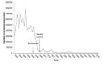
I saw a graph a couple of months ago that shows a huge plunge in measles cases and deaths in the few years leading up to the 1968 licensure of the measles vaccine. The graph actually comes up quickly if you do an image search for “measles graph.” I’ve seen it used to support the argument that the measles vaccine just doesn’t work and is therefore not worth the trouble.

I dug up the primary data used for this graph and made my own (below). It shows that measles cases dropped before introduction of the 1968 vaccine (measles deaths follows the same pattern). But you’ll also see that this plunge occurred two years after the first version of the measles vaccine was licensed in 1963.  I am not a public health expert, but it’s pretty clear that the drop in cases and deaths from measles came after licensure of the initial measles vaccine.

1

I did this research to both appease my curiosity and demonstrate that the question of whether the vaccine works on a population scale has been settled for a long time.  That doesn’t mean that on an individual level it’s impossible to get sick even if being vaccinated against measles.

There are many potential reasons one may not respond to a vaccine. These range from very basic reasons, like the vaccine being stored at the wrong temperature or being injected incorrectly, to complex biological reasons that are active areas of ongoing research.  For example, we know that mothers’ antibodies (or immunoglobulins) circulate in newborns for about 12 months. Although these antibodies can protect a newborn from pretty much anything its mom is immune to, they also prevent the child’s own immune system from generating memory responses to the same diseases. Mom’s antibodies can actually cover up viruses and bacteria and hide them from baby’s immune cells. They can also form complexes with bits of virus or bacteria that bind a receptor that shuts down B cells, the cell responsible for making new antibodies. This receptor, called FcγRIIb, is part of a negative feedback system that tells B cells when they’ve made enough antibodies.

All of these possibilities are part of why the CDC recommends two shots separated by at least a month; statistically, getting two shots means you’re covered because the chance that your immune system would miss out twice in a row is very low. Still, the only way to be 100% sure an individual responded to the vaccine is to measure the level of antibodies in the blood that can bind to and neutralize the measles virus. For different viruses, there are different thresholds for the concentration of antibodies needed to protect a person from getting sick. When a vaccine is first tested, antibody levels are monitored to make sure it actually works. After that, it would be too expensive and unnecessary to measure antibodies in every person who receives the vaccine. So, even though two shots should do the trick statistically, there’s always a remote chance the vaccine just didn’t induce a good immune response, and most of the time, no one would be the wiser.

There was such case in New York City back in 2011, in which a woman who had gotten both requisite vaccinations still managed to get sick. The researchers who did this study measured measles-neutralizing antibodies in the woman’s blood and found that she was making a kind of antibody that the body mainly produces the very first time it sees a pathogen. That kind of antibody is called immunoglobulin M (IgM), and it’s a sort of knee-jerk, immature antibody response that keeps the virus at bay as the immune system generates the more mature, “stickier” IgG. So her body was acting as though it had never seen the measles before, even though she was vaccinated twice.  This is not a huge surprise; even after two shots, the failure rate for the measles vaccine is 3%.

What was unique was that she also ended up spreading measles to 4 other people who were also vaccinated (that’s 4 out of a total of 88 contacts).  All of these people made strong IgG responses and none of them spread the virus to anyone else. And, unlike the first case, none of them were hospitalized—even one who was on immunosuppressive drugs. So this study shows that yes, it is possible to get measles and even spread it to others if you’ve been fully vaccinated. It’s an anomaly, but it’s possible. In fact, that’s what made this case so interesting—the fact that it was so unlikely.

With all the variables of human life, it’s a wonder that the measles vaccine works 97% of the time. This study shows that even when it doesn’t work, the measles virus can’t go far if enough people are vaccinated. The strong secondary immune responses of those 4 cases made their illness less severe and reduced their chances of spreading the disease any further.

Sources

Committee to Review Adverse Effects of Vaccines; Institute of Medicine; Stratton K, Ford A, Rusch E, et al., editors. Adverse Effects of Vaccines: Evidence and Causality. Washington (DC): National Academies Press (US); 2011 Aug 25. 4, Measles, Mumps, and Rubella Vaccine. Available from: http://www.ncbi.nlm.nih.gov/books/NBK190025/

Niewiesk S. (2014). Maternal Antibodies: Clinical Significance, Mechanism of Interference with Immune Responses, and Possible Vaccination Strategies, Frontiers in Immunology, 5 DOI: http://dx.doi.org/10.3389/fimmu.2014.00446

Rosen J.B., C. J. Hickman, S. B. Sowers, S. Mercader, P. A. Rota, W. J. Bellini, A. J. Huang, M. K. Doll, J. R. Zucker & C. M. Zimmerman & (2014). Outbreak of Measles Among Persons With Prior Evidence of Immunity, New York City, 2011, Clinical Infectious Diseases, 58 (9) 1205-1210. DOI: http://dx.doi.org/10.1093/cid/ciu105


2 Comments

A risk worth taking–And one your immune system is prepared to take.

When was the last time you made an important decision with 100% certainty?

Most, if not all, decisions in life come with risks, consequences or trade-offs. Healthcare is no different from anything else. Every surgery, pill, shot, even every new diet or exercise routine has its risks. And vaccines are not exempt. It’s true, vaccines have risks (probably the most common one for most vaccines is soreness at the injection site). And it’s no secret either—check out this list on the Center for Disease Control’s (CDC) website. They even list extremely rare reported events that they can’t prove were related to vaccination, but occurred around the same time.

Early recipients of vaccines understood side effects all too well. In the 1700’s, vaccination against smallpox, which entailed rubbing pus from an afflicted person into a small cut, was known to cause a mild form of disease, and in 1-2% of cases, death. But to those who saw what real smallpox could do firsthand, the risk was worth it, because even if they didn’t yet know how it worked, they knew that vaccination saved lives.

These days, vaccines are far safer, but the fear of potential side effects often overshadows the fear of disease. Perhaps the most notorious of these fears is the alleged and debunked link between autism and the measles, mumps and rubella (MMR) vaccine.  Many researchers have taken an honest and thorough look at this and the question has been settled from a scientific standpoint.

As is the case with everything, though, people factor things besides scientific evidence into their decisions. For example, a sense of social responsibility may influence your decision to get the flu shot each year. You may also factor in anecdotes about a co-worker’s friend getting the flu after being vaccinated. Though rejecting one piece of information and blindly accepting another is everyone’s right, making an informed decision requires consideration of all types of information.

Many take the reasonable route of deferring to their doctors who have hopefully kept abreast of the scientific evidence and have likely seen the anecdotal evidence first hand. A doctor may defer to the recommendations of an organization like the Advisory Committee on Immunization Practices (ACIP), a rotating group of doctors and scientists who painstakingly study the science and side effects of every vaccine that goes onto the market. You can learn more about ACIP here and even attend their meetings if you want.

Then there are some who would like to have a couple of questions answered and to feel more involved and informed about their own, or their children’s health care. And then some who are just plain scared of the potential side effects. These lingering questions and fears surrounding vaccination are worth addressing (not to mention scientifically fascinating). For a thorough list of such questions, I recommend this site (and of course, it’s always wise to speak with a trusted healthcare professional about your concerns). Over the next couple of posts, I plan to explore some recent research that sheds light on a just couple of these questions:

First, can a vaccine make you sick? And second, why do vaccinated people still catch disease?

One way to explore the first question is look at the differences between the altered form of a virus found in a vaccine and the real deal. For something like the flu shot, which contains dead virus, the difference is obvious. If the virus is not alive, it can’t get into cells and replicate. It can, and should, activate immune cells, which could bring along soreness or a headache.


You may have heard about people getting the flu, or flu symptoms from the flu shot itself. There is some evidence that the act of getting the flu shot can put you at risk for the flu. One study published last year concluded that just going to the doctor slightly increased the probability of experiencing flu-like symptoms within the following two weeks (read this for more). If you get the vaccine at a clinic or doctor’s office, you could increase your chance of contact with people who have the flu or surfaces they have recently touched. It takes about a week for your body to generate antibodies good enough to protect you from the virus, so it’s definitely possible to get sick just after being vaccinated. For more flu myths, check out this list.


For some diseases, like measles, the immune system really needs to see a live vaccine to generate long-term immunity. The reason for this is not completely clear, but we do know that it takes a while for our bodies to generate the “best and brightest” long-lived immune cells and a dead vaccine may be cleared too quickly for this to happen. So, we’re stuck with live vaccines, at least until researchers come up with something better.

Do live vaccines have more risks than dead ones? Well, for some people, yes. There are a handful of case reports of kids with rare genetic immunodeficiency disorders getting polio from the vaccine, and live vaccines could make someone with uncontrolled AIDs sick. However, there have been very few reports of HIV+ people getting sick after receiving a live viral vaccine (Summarized here).  And just to be safe, the CDC recommends pregnant women and those on immune-depleting chemotherapy avoid most live vaccines, though there is not a lot of data for or against them in those cases.

Measles pneumonia - Histopathology

Lung cells fusing together into one, measles-infected “giant cell.”

But what about in the average healthy person? What happens after a live virus vaccine enters your body, and how is it different from a live, natural, infectious virus? Let’s take a closer look at the recently popular measles vaccine. The virus used in for measles vaccine is “weakened” because it’s been grown, harvested, and grown again and again in human or chicken cells in culture dishes. The virus adapted to its environment in a culture dish, and lost its potency in the human body. On a molecular scale, scientists are still collecting information about exactly how this “weakening” happens. One thing they know is that the vaccine version of the virus infects different kinds of cells than the natural version of the virus does.

One researcher working toward a better understanding of this question is W. Paul Duprex, at the Boston University School of Medicine. His lab engineered measles viruses to glow by giving them the gene for the jellyfish green fluorescent protein (GFP). Then they infected macaques monkeys with either the infectious natural measles virus or the vaccine strain and looked for the glowing viruses in different parts of the animals’ bodies. When they looked for the virus in blood or throat swabs, they found much less—orders of magnitudes less—of the vaccine strain compared to how much natural virus was growing in the monkeys. The researchers also examined slices of lymph nodes with a microscope and measured GFP in immune cells using a laser and detected very little, if any, of the vaccine virus strain inside immune cells. The infectious version, on the other hand, seemed to love infecting and dividing inside of immune cells.

Both viruses were able to infect one type of innate immune cell, but only in the lungs. And, it’s important to note that the scientists delivered both types of virus straight into the animals’ airways, so both strains had ample opportunity to infect. Just this month, though they published a study that mimicked the actual vaccine route, which is an injection into a muscle, and saw that the vaccine virus also only infected innate immune cells in the muscle. To see pictures of Duprex’s “glowing” virus infecting these cells, check out this recent National Geographic blog post.

When these innate immune cells, called dendritic cells and macrophages, get infected, they display little bits of the virus to other immune cells in nearby lymph nodes. For this reason, they are called professional antigen presenting cells. Other immune cells in the lymph nodes will generate a response, clear the present virus, and remember it well enough to prevent infection with the natural version in the future.

If innate immune cells brought the natural virus to the lymph nodes, cells in the lymph node would become infected and the virus would continue to spread throughout the body. This research is just getting started, but so far it looks like the vaccine version of the virus is well contained by dendritic cells and macrophages. They are professionals after all, and they do this kind of thing all day every day.

So, should you fear live viral vaccines? Well, do you fear the live bacteria, viruses and fungi living all over your body? Your immune system has done a good job at keeping them in check so far. If you’re generally healthy, a live viral vaccine is like a blip on your immune system’s radar.

I think of it like going on a roller coaster. You can stand in line and mull over all of the things that have a one in a million chances of going wrong, or consider the actual data–the hundreds of people who rode it without any incident just during the time you were in line.

In the case of live vaccines, millions of people have had them with no incident just in the past year. And unlike a roller coaster ride, the marginal risks of measles vaccination are exchanged for a major, life-long benefit.

Please note:

I am not a medical professional and the opinions within this blog are not intended to be used as medical advice.

Sources:

Gerber J. (2009). Vaccines and Autism: A Tale of Shifting Hypotheses, Clinical Infectious Diseases, 48 (4) 456-461. DOI: http://dx.doi.org/10.1086/596476

Simmering J.E., Joseph E. Cavanaugh & Philip M. Polgreen (2014). Are Well-Child Visits a Risk Factor for Subsequent Influenza-Like Illness Visits?, Infection Control and Hospital Epidemiology, 35 (3) 251-256. DOI: http://dx.doi.org/10.1086/675281

Angel J. (1998). Vaccine-Associated Measles Pneumonitis in an Adult with AIDS, Annals of Internal Medicine, 129 (2) 104-106. DOI: 10.7326/0003-4819-129-2-199807150-00007

de Vries R.D., Lemon K., Ludlow M., McQuaid S., Yüksel S., van Amerongen G., Rennick L.J., Rima B.K., Osterhaus A.D.M.E. & de Swart R.L. & (2010). In vivo tropism of attenuated and pathogenic measles virus expressing green fluorescent protein in macaques., Journal of virology, PMID: http://www.ncbi.nlm.nih.gov/pubmed/20181691

Rennick L.J., Thomas J. Carsillo, Ken Lemon, Geert van Amerongen, Martin Ludlow, D. Tien Nguyen, Selma Yüksel, R. Joyce Verburgh, Paula Haddock & Stephen McQuaid & (2014). Live-Attenuated Measles Virus Vaccine Targets Dendritic Cells and Macrophages in Muscle of Nonhuman Primates, Journal of Virology, 89 (4) 2192-2200. DOI: http://dx.doi.org/10.1128/jvi.02924-14


Leave a comment

The most promising treatments for ebola are based on basic immunology-part 2

One of the more peculiar, historic and almost cinematic treatments being discussed in the midst of the ebola crises is the use of blood transfusions. In movies, the blood of a survivor or someone special is often supposed to have some sort of mystical effect on the (usually villainous) recipient. It turns out, blood transfusions from people who have survived ebola are nearly as mystical.

It seemed obvious to me at first that the active components in blood transfusions from ebola survivors must be anti-ebola antibodies. Such antibodies would neutralize the virus and help the immune system clear it out. And in fact, a 1999 study reported that seven patients who survived the 1995 outbreak in the Democratic Republic of the Congo after receiving transfusions from survivors had anti-ebola antibodies circulating in their blood. One kind of antibody, called IgM, was absent in another patient who received a transfusion and died. This very small study seemed to indicate that transfusions could work against ebola and that antibodies are key to making them work. (You may be wondering why, if the transfusions worked, more patients weren’t treated this way during the 1995 outbreak. One important reason is that that blood cannot be transfused unless the donor and recipient blood types are compatible.  So the treatment is limited by the number of willing survivors and their blood types.)

This idea was challenged by a 2007 study done in a nonhuman primate species, rhesus macaques. For this study, researchers drew blood from a rare few monkeys who had survived an ebola infection four or five years earlier and a second “boost” infection 30 days earlier. They transferred the blood into other, recently-infected animals, and none of them survived…even those that made a lot of antibody. There are, of course, caveats. Monkeys are not humans, after all. It is possible that they fight the virus differently. And in the discussion of the paper, the researchers admit the experiments that had successfully transferred antibody-mediated immunity in guinea pigs had not worked in rhesus macaques.

There are also caveats to the human study though. The main one being that it can’t account for the better treatment transfusion patients received compared to other patients. The seven may have survived simply because they received better care in the clinics. The boost of cells, fluids, proteins and electrolytes that come along with blood transfusions may also have helped.

Scanning electron micrograph of red and white blood cells (National Cancer Institute)

Scanning electron micrograph of red and white blood cells (National Cancer Institute)

In spite of it all, the World Health Organization is behind blood transfusions and transfer of plasma from ebola survivors.  Plasma is the liquid part of blood that contains proteins like antibodies, along with other things like electrolytes and hormones. The first of the eight original transfusion patients, a 27 year-old nurse, was originally supposed to receive plasma and not whole blood. This was because, in 1978, a researcher who received plasma survived an ebola infection brought on by a finger prick in the lab. The nurses’ doctors settled for blood because they didn’t have the right tools to separate the plasma (a process called plasmapheresis).

The seven transfusion patients who followed the nurse ranged from a 54 year-old woman to a 12 year-old girl who caught the virus by kissing her newborn niece just days before the infant died. Antibodies seem to be the most likely explanation for the high rate of survival, but it is still not clear whether they were. Well-controlled human trials to determine whether blood transfusions work for ebola will probably never be possible. But, more and more people may be receiving them, so there may soon be more information about whether and how they work.


Leave a comment

The most promising ebola treatments are based on basic immunology

Though for many of us, the ebola crises is oceans away, the epidemic still weighs heavily on the hearts and minds of people all over the world. For some researchers, public health officials and drug developers, it is the driving force of all daily activity. Right now, there are two vaccines and eight treatments being developed or tested for their effectiveness against controlling infection or stopping the  virus’ spread. The most encouraging results have come from treatments that rely on a very basic aspect of immunology: antibodies neutralize viruses.

Antibodies are proteins made by immune cells called B cells. Each one of your millions of B cells is capable of producing antibodies specific for one thing, and when a B cell comes into contact with that one thing, it secretes lots of antibodies. The antibodies then tag invading pathogens, like viruses, to make other immune cells aware of the invader’s presence. If enough antibodies stick to a virus, they can cover it up, or neutralize it, and prevent it from infecting cells.

Ebola infection does trigger an antibody response, but for reasons that are still being studied, those antibodies are not usually enough to stop the virus before it spreads throughout the body. The concept behind ebola treatments like Zmapp, blood transfusions, vaccines and even supportive care, is to help the immune system outpace the growing virus.


Zmapp

Over the summer, this product was on headlines everywhere. Zmapp sparked a controversy over who should get the most cutting-edge treatments when it was given to two missionary doctors who flown to Atlanta for care.  Zmapp is not really a drug; it’s a combination of three kinds of antibodies that bind to the surface of ebola virus particles. Because each type was originally produced by one individual B cell they are called monoclonal antibodies. Monoclonal antibodies are used for treating cancer, autoimmune diseases and other infections.

Identifying the right monoclonal antibodies can be a painstaking and years-long process. Researchers collect B cells from a person or animal in the midst of an active immune response, in this case, against ebola. Then they seed individual antibody-making B cells into tiny wells on a cell culture dish. Later they test the culture media from each well for the presence of antibodies and select the cells making the best antibody to be “immortalized.” Cells are immortalized by altering their genes or fusing them with cancer cells that are already immortal. Usually the cells are frozen and stored for later use. They can be thawed anytime and grown to large quantities to make antibody.

Forget cigarettes...tobacco plants have lots of potential for "pharming" biological drugs like the monoclonal antibodies in Zmapp (From Wikemedia Commons)

Forget cigarettes…tobacco plants have lots of potential for “pharming” biological drugs like the monoclonal antibodies in Zmapp (From Wikemedia Commons)

It seems simple, but getting the process right can take years. The monoclonal antibodies in Zmapp were originally derived in mice back in 2000.

From there, the antibodies have to be purified. It can take liters and liters of cell media to purify enough antibody to treat one person one time. As a therapeutic, monoclonal antibodies are typically dosed over multiple treatments. In a recently published study showing the effectiveness of Zmapp against ebola infected monkeys, the animals were treated three to five times a day.

There are alternative ways to do this, however. Because antibodies are proteins they are coded by specific genes. So instead of fusing selected B cells with cancer cells, researchers could copy the gene coding for the cell’s antibody and put it into something else, like bacteria or, in the case of Zmapp, tobacco plants. Many biological products, like insulin have been produced in bacteria since the 1980s. Plant production of human proteins is a bit more recent.  The first human protein produced in plants in 2012 for a medical purpose was an enzyme injected into patients who can’t make it themselves. Some insulin is also now produced in plants.

Unlike cell-based or bacteria-based approaches, plants don’t have to be genetically manipulated and then grown up and harvested. Instead, adult plants are infected with viruses engineered to express the antibody-coding genes. The viruses introduce the genes, and the plants make the antibody. For some proteins, this results in much higher yields than cell-based methods. The monoclonal antibodies in Zmapp are being made by three different companies using a variety of these methods.

But there is a catch. When any kind of cell (plant, animal, bacteria) produces a protein, it adds little sugar labels to keep track of it during each stage of production. This process is called glycosylation. These glyo-labels vary by species and they can affect the way a protein functions. Because of this, the plants being used to grow Zmapp are not your run-of-mill tobacco. They are genetically modified so that they can give the anti-ebola antibodies more human-looking labels. That adds another layer of complexity to be addressed as these companies start to make large quantities of Zmapp.

It’s fascinating how this technology was developed step by step—often in obscurity—over the course of many decades.  Hopefully, it will be scaled up successfully in the coming months to provide more much-needed doses.

Next time…Blood transfusions.


Leave a comment

Banking on baby: all about umbilical cord blood stem cell transplants

Over the summer, my then-pregnant friend asked for my opinion about umbilical cord blood banking, naturally sending me into a world of fascinating biology, cutting edge medicine and some ethical quandaries.

If you can afford the $1000-2000 processing fee and at least $100 a year to store the blood, banking seems like a no-brainer. “You never know,” rings in the backs of many expecting parents’ minds as the one-time opportunity approaches. But there is more to consider than price. The biology behind the technique and the currently available applications of frozen cord blood may influence one’s decision about whether to bank, and also how and where to do it.

Cord blood contains a high frequency of hematopoietic stem cells, which can differentiate into any kind of blood cell. They can mature into megakaryocytes that make platelets, red blood cells, or immune cells like B cells or eosinophils. We all carry these stem cells throughout our lives, mainly in our bone marrow, and they produce cells that periodically replace blood cell populations.

Blood cells arising from hematopoietic stem cells. (Wikimedia commons, based on original by A. Rad)

Blood cells arising from hematopoietic stem cells. (Wikimedia commons, based on original by A. Rad)

Cancers rising from white blood cells (like lymphoma or leukemia) and genetic defects interfering with the production of any kind of blood cell can conceivably be addressed by resetting the whole system with a bone marrow (or stem cell) transplant.  Transplants using donated bone marrow have been used to do just this for about 50 years. In the late 1980s, it became clear that cord blood stem cells could do the same with some distinct advantages.

For one thing, collecting cord blood is much less difficult and invasive compared to harvesting bone marrow. Bone marrow donation involves anesthesia and a very large needle stuck directly into the bone. Stem or progenitor cells can also be separated from adult blood through a process called apheresis, but it is no cakewalk, especially compared to harvesting cord blood, which simply involves injecting a needle into the cord after it’s been cut.

Donor matching is also more flexible for cord blood. To avoid graft rejection, stem cell (and all organ) donors and recipients are matched for proteins expressed on the surface of immune cells. If they don’t match, the T cells in the donated transplant may attack the tissues of the recipient. T cells found in cord blood respond with less gusto and there are higher frequencies of T cell subsets that control the immune response called T regulatory cells. This means there’s a lower chance of the donor immune system harming the recipient.

Given these advantages, is banking worthwhile? It depends on what you hope to get out of it. When you think about storing a baby’s cord blood, you may think it’s for the sake of that particular child. The truth is, the stem cells in that kid’s blood are more likely to be useful for someone else. That was the case of the first ever cord blood transplant performed in 1988. A five year-old boy with a rare genetic disease called Fanconi Anemia received cord blood cells from his newborn sister. At the time, the boy’s white blood cell counts were dropping because inherited defects in a DNA repair pathway made it impossible for his bone marrow to produce healthy blood cells quickly enough. His own cord blood, of course, would have been useless because the same genetic defect would manifest again. Today, that patient is a grown man, but he has a female blood and immune system thanks to his sister.

Cases covered in the news about cancer patients cured because of cord blood transplants are usually about patients who received donated cord blood from public banks. In fact, their own stem cells would not have worked. In such cases, the transplant is an imperfect match on purpose so that the new immune system will attack cancer cells that the old immune system was blind to. This is typically done for blood cancers like leukemia. A transplant of a close or perfect match is desirable for patients whose bone marrow is depleted as a side effect of chemotherapy and/or irradiation for other types of cancer. However, stem cells for this kind of transplant can also be harvested from one’s own bone marrow or blood before beginning treatment.

medium_7413610060

photo credit: Banc de Sang i Teixits via photopin

Even if the chances of using one’s own cord blood are remote, it may be desirable to store it in case a family member could use it. There are a couple of caveats to consider, however, before committing to private banking. First, it’s been estimated that at least 70% of adult recipients need two units of cord blood to successfully reinstate a new blood and immune systems That means it’s likely that even if you do save your baby’s cord blood, it may not be enough if he or she needs it as an adult. This may change in the future; a study published last week in Science found that a drug compound called UM171 kept human cord blood stem cells “immature” while allowing them to expand. There are also several clinical trials underway that will test whether expanding cord blood progenitors through other means can reduce the number of units needed and increase transplant success.

The second caveat is a little bit more about logistics and politics than science. Right now, only 10% of collected cord blood meets the standards required for transplantation. These standards included how many cells are present, how many cells survived and whether the blood was collected, shipped and frozen properly. (For an interesting glimpse at what could go wrong, check out this Wall Street Journal article).  And the way cord blood units are handled and stored is only regulated by the Food and Drug Administration if they are stored in public banks. That is not to say that there are no good and reputable private banks. It is, however, important to recognize that private banking requires lots of research and care when choosing a company.

I mentioned that the chances of performing a cord blood transplant on the original donor are not very high given the current uses of cord blood stem cells—mainly to replace blood stem cells in the bone marrow. That is not the whole story though. There are clinical trials going on to test the therapeutic effects of cord blood stem cells for things like cerebral palsy, type I diabetes and even hearing loss. These studies are based on observations suggesting stem cells found in cord blood can reduce brain damage after injury, but it’s not yet clear how. There are also other kinds of stem cells in cord blood that can differentiate into cells other than blood cells (pancreatic cells for example). There may still be a lot of untapped potential for cord blood. For many parents, that is enough reason to put their kids’ blood “on ice” and wait it out.

Sources:

Metheny L., Caimi P. & de Lima M. (2013). Cord Blood Transplantation: Can We Make it Better?, Frontiers in oncology, PMID: http://www.ncbi.nlm.nih.gov/pubmed/24062989

Gluckman E., Arleen D. Auerbach, Henry S. Friedman, Gordon W. Douglas, Agnès Devergie, Hélène Esperou, Dominique Thierry, Gérard Socie, Pierre Lehn & Scott Cooper & (1989). Hematopoietic Reconstitution in a Patient with Fanconi’s Anemia by Means of Umbilical-Cord Blood from an HLA-Identical Sibling, New England Journal of Medicine, 321 (17) 1174-1178. DOI: http://dx.doi.org/10.1056/nejm198910263211707

Wagner J.E. Should double cord blood transplants be the preferred choice when a sibling donor is unavailable?, Best practice & research. Clinical haematology, PMID: http://www.ncbi.nlm.nih.gov/pubmed/19959107

Fares I., Chagraoui J., Gareau Y., Gingras S., Ruel R., Mayotte N., Csaszar E., Knapp D.J.H.F., Miller P. & Ngom M. & Cord blood expansion. Pyrimidoindole derivatives are agonists of human hematopoietic stem cell self-renewal., Science (New York, N.Y.), PMID: http://www.ncbi.nlm.nih.gov/pubmed/25237102

Petrini C. (2014). Umbilical cord blood banking: from personal donation to international public registries to global bioeconomy., Journal of blood medicine, PMID: http://www.ncbi.nlm.nih.gov/pubmed/24971040


Leave a comment

Vaccination Coverage and Herd Immunity

Great info about how herd immunity works and how important it is to consider how health choices that seem purely personal can affect others.

Parasite Ecology

I’ve talked about vaccination and herd immunity on this blog before, but I think it’s important for me to emphasize how INCREDIBLY IMPORTANT it is to get vaccinated.  The importance of vaccinating most of the population is usually explained using mathematics, because scientists study the spread of pathogens by using mathematics.  But today, I’m going to try to explain it with cartoons and pictures, instead of math.

herdimmunity1

herdimmunity2

Without explaining the math, I’ll say that there are some “magic numbers” for vaccination.  These numbers are unique to each pathogen/disease.  For instance, for whooping cough, a disease that can make make babies very sick, the “magic number” is between 92 and 94.  That is, 92-94% of people must be vaccinated in order to prevent disease epidemics of whooping cough.  If that magic number – called the herd immunity threshold – is reached, babies are indirectly protected from whooping cough.  If not…

View original post 233 more words


1 Comment

Allergies are no fun…but the biology behind them is!

Spring is nearly upon us and along with trees and flowers, seasonal allergies will bloom once again.  Even though allergies can be annoying, debilitating and even life-threatening, the science behind them is fascinating.  Science published a timely paper at the end of February describing some of the ways different kinds of allergens work.  Allergens are small parts—individual proteins or molecules—of things that cause allergic responses.

The group who published the study worked with cells called mast cells, one of the common types of the immune cells that respond to allergens and make you itchy, sneezy and swollen.  Before they can activate mast cells, allergens have to be recognized by a particular type of antibody, or immunoglobulin, called immunoglobulin E, or IgE.  On one end, IgE binds an allergen, and on the other it interacts with a protein receptor on mast cell surface.

By connecting the mast cell to the allergen, IgE gives the mast cell permission to do its thing, and its thing is called degranulation.  Mast cells are brimming with packets, or granules, of histamine and heparin and other proteins that damage microbes as well as tissue.  When the cells degranulate, they open up and release their contents into whatever tissue they happen to be in—the skin, the lungs or the gut for example.  Many of the contents released make blood vessels leaky and attract lots of immune cells, causing inflammation.  Antihistamines prevent the released histamine from binding its receptors on blood vessel cells.  Another treatment option currently under investigation is a drug that blocks the interaction between IgE and the receptor on mast cells to prevent this process from even getting started.  

The recent Science paper took a close look at the mast cell response to IgE-bound allergen and showed just how fine-tuned it can be. The researchers activated mast cells with allergens that bound tightly or weakly to IgE and found that the strength of the interaction, also called affinity, changed the way that mast cells responded.

http://commons.wikimedia.org/wiki/File%3ASMCpolyhydroxysmall.jpg

Skin mast cells stained with Toluidine blue

The researchers could study mouse mast cells in culture dishes, because mast cells grow up from stem cells inside bone marrow.  So they grew up mast cells from mouse bone marrow and then gave them the strongly binding allergen (high affinity) or the weakly binding one (low affinity). They could get the mast cells to respond and degranulate with both, but it took 100 times more of the weak binding allergen to get the same response caused by the strong one.

To understand how allergic reactions work in living creatures, researchers often sensitize mouse ears by exposing them to an allergen and later re-introduce the allergen through the bloodstream. Then they can measure how inflamed the ears get and how many and what kinds of immune cells travel to the ear after injecting the allergen.  In this study, the strong binding allergen caused more intense and more sudden ear inflammation and immune cell infiltration than the weaker binding allergen.

So how does this fascinating mechanism actually relate to human allergies, which for some people is a life-threatening condition.  Although some allergies go away with age, there is currently no permanent cure for those that don’t.  Treatment of serious allergies is centered around desensitization immunotherapy, which is just repeated exposure to small doses of allergen over time.  The treatment may last anywhere from months to a lifetime and there are no biomarkers, or biological tests, that tell doctors when the treatment is working.  Instead, they simply test allergens on patients, which could mean pricking the skin or making them eat peanuts one at a time until they do or don’t get sick.  

A clinical study that came out in January helped me understand how knowledge of allergen binding strength could be helpful in treatment.  In this study, children with milk allergies were undergoing oral immunotherapy, which in this case simply meant they had to drink small amounts of milk that were increased over time.  The researchers collected serum samples from the kids in the study and measured levels of IgE as well as the affinity of IgE for proteins found in cow’s milk to see if either would change as kids became more tolerant to milk.

In some cases, the immunotherapy had to be discontinued because the reactions to milk were too severe.  The researchers found that the IgE from the kids whose treatment was discontinued bound more tightly to milk proteins compared to kids who responded well to the treatment.  So the strength of the interaction between IgE and allergens does matter, at least in the case of cow’s milk allergies. This study didn’t look at mast cells, but it does indicate that the molecular details of how IgE connects allergens to mast cells are worth studying.  Those details can provide clues about what is going on inside a person with allergies and how well they may respond to immunotherapy.

Sources:

Mastcellaware.com (A whole website about Mast Cells)

Suzuki R., Leach S., Liu W., Ralston E., Scheffel J., Zhang W., Lowell C.A. & Rivera J. (2014). Molecular Editing of Cellular Responses by the High-Affinity Receptor for IgE, Science, 343 (6174) 1021-1025. DOI:

Savilahti E.M., Kuitunen M., Valori M., Rantanen V., Bardina L., Gimenez G., Mäkelä M.J., Hautaniemi S., Savilahti E. & Sampson H.A. & (2014). Changes in IgE and IgG4 epitope binding profiles associated with the outcome of oral immunotherapy in cow’s milk allergy, Pediatric Allergy and Immunology, n/a-n/a. DOI:

Moran T.P., Vickery B.P. & Burks A.W. (2013). Oral and sublingual immunotherapy for food allergy: current progress and future directions, Current Opinion in Immunology, 25 (6) 781-787. DOI:


Leave a comment >

Most of us have had a few run-ins with the bane of the kitchen, the fruit fly.  Known better by some as Drosophila melanogaster, scientists have been studying the species since the early 1900s. But until college labs, I never understood what you would want to know about them other than how best to keep them out of your bananas. 

Believe it or not, research using fruit flies has given us much of our modern understanding of genetics. Research using fruit flies led to the idea that chromosomes contain genes, the concept of mapping where genes are within a chromosome, the fact that ionizing radiation causes genetic mutations and the fact that XX chromosomes makes females female and XY makes males male.

By André Karwath aka Aka (Own work) [CC-BY-SA-2.5 (http://creativecommons.org/licenses/by-sa/2.5)], via Wikimedia Commons

Drosophila melanogaster

Fruit flies have more recently been used to understand human immunology.  A study published last month in PlosOne describes immune responses of flies that were born in outer space aboard the shuttle, Discovery.  It may sound far-fetched, but fruit flies are a cheap and compact model organism that has a lot in common with humans, especially when it comes to immunity.

I’ve written a lot about the adaptive immune system—mainly B cells and T cells with very specific receptors that recognize very specific parts of infecting pathogens and become memory B and T cells.  There is another branch of immune cells that make up the innate immune system.  They have receptors that detect general patterns found on pathogens, called (appropriately) pattern recognition receptors.  For example, some recognize sugars arranged in repeated patterns on the surface of bacteria.  Pattern recognition receptors are less specific than receptors on B and T cells, and don’t generate memory.  Instead, they act as a first line of defense that allows immune cells to react quickly and make signaling proteins to direct the immune response toward inflammation.

Toll-like receptors make up the class of innate receptors that kicked off studies into the innate branch of immunity. They were given their name due to their shared genetic sequence with a fruit fly receptor called Toll.  Studies in fruit flies gave researchers major clues throughout the early 1990’s about how Toll-like receptors work. (For a complete history of how it all happened, check out this Nature review, or this free article.)  Toll-like receptors are found on both adaptive and innate immune cells, as well as neurons and epithelial cells. We are constantly sensing our environment via Toll-like receptors. They are vital for responding to viral and bacterial infections and help keep bacteria in our gut microbiomes under control.  Many researchers are looking for the best ways to activate them during vaccination in place of current adjuvants.  It may seem odd, and maybe a little bit gross, but without those pesky flies, we would not know nearly as much about how our own immune systems function.

In case you were curious, the flies born on the space shuttle Discovery—called “space flies” by the authors of the paper—were infected with fungal spores and bacteria and the researchers examined changes in genes involved in immune responses. Unfortunately, the “space flies” were overall immunosuppressed and at a disadvantage compared to “earth flies,” leading the authors  to conclude that gravity plays a key role in immune function.

Sources:

Medzhitov R. & Janeway C.A. (1998). Self-defense: The fruit fly style, Proceedings of the National Academy of Sciences, 95 (2) 429-430. DOI:

O’Neill L.A.J., Golenbock D. & Bowie A.G. (2013). The history of Toll-like receptors — redefining innate immunity, Nature Reviews Immunology, 13 (6) 453-460. DOI:

Taylor K., Kleinhesselink K., George M.D., Morgan R., Smallwood T., Hammonds A.S., Fuller P.M., Saelao P., Alley J. & Gibbs A.G. & (2014). Toll Mediated Infection Response Is Altered by Gravity and Spaceflight in Drosophila, PLoS ONE, 9 (1) e86485. DOI:

http://web.mit.edu/HST.160/www/DrosophilaGenomeResearch.pdf

http://www.benchfly.com/blog/model-organism-week-drosophila-melanogaster-the-fruit-fly/